![]() Figure 4 of
Maddala, Mol Vis 2003;
9:329-336.
Figure 4 of
Maddala, Mol Vis 2003;
9:329-336.
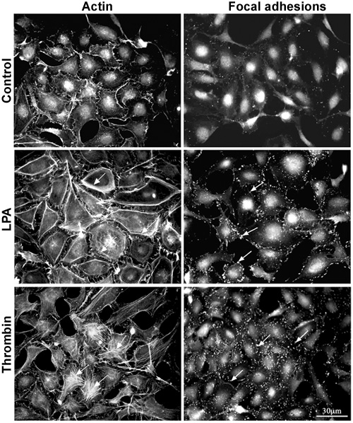
Figure 4. LPA and Thrombin induced cytoskeletal changes
Human lens epithelial cells maintained in 1% serum for 24 h were treated for 1 h with either LPA (5 μg/ml) or thrombin (1 Unit/ml) in the absence of serum, then fixed and stained for detection of actin stress fibers and focal adhesions, as described in methods. LPA and thrombin both induced formation of actin stress fibers (indicated with arrows in the left panel) and focal adhesions (indicated with arrows in the right panel), however, as compared to thrombin, LPA induced very intense cortical stress fibers whereas thrombin caused formation of transverse actin stress fibers. LPA treated cells with intense cortical actin stress fibers retracted from each other compared to controls and to thrombin treated cells. As in the case of TGF-β, FGF, and PDGF, increased actin stress fibers are associated with increased focal adhesions at leading edges of the cell membrane in LPA treated cells.