![]() Figure 3 of
Maddala, Mol Vis 2003;
9:329-336.
Figure 3 of
Maddala, Mol Vis 2003;
9:329-336.
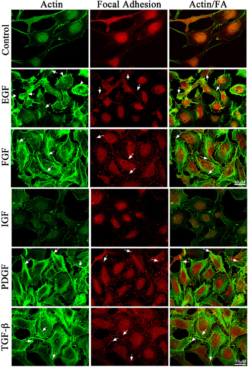
Figure 3. Growth factor-induced actomyosin reorganization and focal adhesion formation
Treatment of semi-confluent cultures of human lens epithelial cells maintained under serum deprivation with different growth factors (at a concentration of 20 ng/ml) led to reorganization of the cellular actin cytoskeleton. Addition of b-FGF, PDGF, and TGF-β induced very strong actin stress fiber formation (FITC-phalloidin staining indicated with arrows), which were localized predominantly to the cortical regions of the cell body indicated with arrows. EGF induced membrane ruffles (indicated with arrow heads) along with weak transverse actin stress fiber formation (indicated with arrows). Stimulation with b-FGF, PDGF, and TGF-β induced very intense focal adhesions, as shown by the arrows (middle panel), whereas EGF caused a marginal increase in focal adhesions compared to untreated controls. Interestingly, unlike other growth factors, addition of IGF-1 caused no obvious change in actin cytoskeletal organization or focal adhesion formation. Images in the third panel from left, depict the costaining of actin stress fibers (green) and focal adhesions (yellowish-orange indicated with arrows) induced by different growth factors. As shown by the arrows, increased actin stress fibers at the leading edges of cell membranes are associated with increased focal adhesion formation. In addition, cells treated with FGF, PDGF, and TGF-β undergo noticeable changes in cell shape, appearing rigid with increased cortical actin stress fiber formation, as compared to untreated cells. Bars indicate the magnification.