![]() Figure 4 of
Fujii, Mol Vis 2003;
9:315-322.
Figure 4 of
Fujii, Mol Vis 2003;
9:315-322.
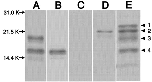
Figure 4. Characterization of antibodies
Characterization of antibody specific to the truncated form of αA-crystallin (1-154). The α-crystallin fraction prepared from bovine lenses was analyzed by western blotting. The blots were stained with the anti-native form of αA-crystallin antibody (lane A), with the anti-truncated form of αA-crystallin antibody (lane B), with the anti-truncated form of αA-crystallin antibody in the presence of 500 μg/ml antigenic peptide (CGVDAGH, lane C), with the anti-native form of αB-crystallin antibody (lane D). Lane E is the Coomassie Brilliant Blue staining pattern of the PVDF membrane. Protein bands: 1, unknown; 2, native form of αB-crystallin (1-175); 3, native form of αA-crystallin (1-173); 4, truncated form of αA-crystallin (1-154).