![]() Figure 5 of
Wentz-Hunter, Mol Vis 2003;
9:308-314.
Figure 5 of
Wentz-Hunter, Mol Vis 2003;
9:308-314.
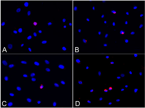
Figure 5. Detection of apoptotic cells
Detection of apoptotic cells through immunofluoresence staining using a monoclonal antibody for ssDNA. Control (A) and DEX-treated (C) corneal fibroblasts contained few apoptotic cells depicted by bright pink nuclear staining. DAPI staining of the nucleus is shown in blue. After exposure to staurosporine, the number of apoptotic cells was increased to a similar extent in control (B) and DEX-treated (D) fibroblasts.