![]() Figure 1 of
Wentz-Hunter, Mol Vis 2003;
9:308-314.
Figure 1 of
Wentz-Hunter, Mol Vis 2003;
9:308-314.
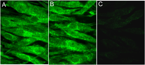
Figure 1. Immunofluorescence staining of myocilin in human corneal fibroblasts
Both untreated control (A) and DEX-treated (B) cells were stained with anti-myocilin. Only background staining was observed when DEX-treated cells were stained with anti-myocilin that was preabsorbed with the synthetic peptide prior to immunostaining (C). Immunostaining with preimmune sera similarly yielded negative results as the preabsorbed antibody (data not shown). The specimens were examined under a Zeiss 100M microscope.