![]() Figure 2 of
Inglehearn, Mol Vis 2003;
9:295-300.
Figure 2 of
Inglehearn, Mol Vis 2003;
9:295-300.
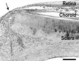
Figure 2. The eye of a blind rge bird at four years of age
Note focal destructive lesion of the retina, choroid and sclera. Away from the lesion (right) the sclera, choroid and retina are intact. At the lesion (arrow) the sclera shows destruction of the cartilaginous plate, the choroid is lost and atrophic and gliotic retina is displaced into the chronically inflamed sclera. Haematoxylin and eosin. Scale bar represents 120 μm.