![]() Figure 1 of
DePianto, Mol Vis 2003;
9:288-294.
Figure 1 of
DePianto, Mol Vis 2003;
9:288-294.
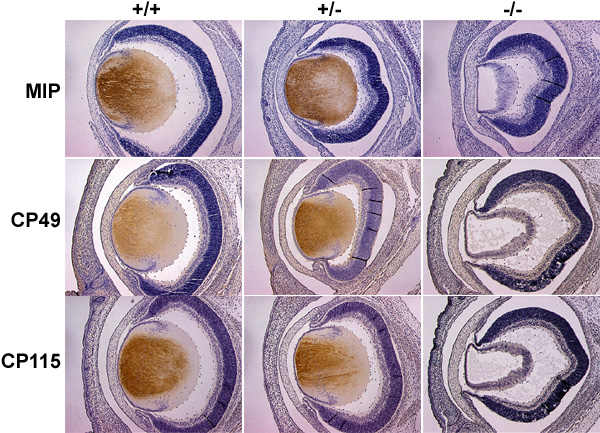
Figure 1. Immunocytochemical localization of MIP, CP49, and CP115 expression
Immunocytochemical localization of MIP, CP49, and CP115 expression in wildtype (+/+), heterozygous mutant c-Maf (+/-), and homozygous mutant c-Maf (-/-) mice. Paraffin sections of E16 to E19 mouse embryonic head were used in immunohistological studies to analyze gene expression. The dark brown color denotes reactivity to the protein of interest. While the lenses of the wildtype and heterozygous mutant c-Maf mice exhibit normal lens development, homozygous c-Maf mutant show no signs of secondary fiber cell formation.