![]() Figure 2 of
Hackam, Mol Vis 2003;
9:262-276.
Figure 2 of
Hackam, Mol Vis 2003;
9:262-276.
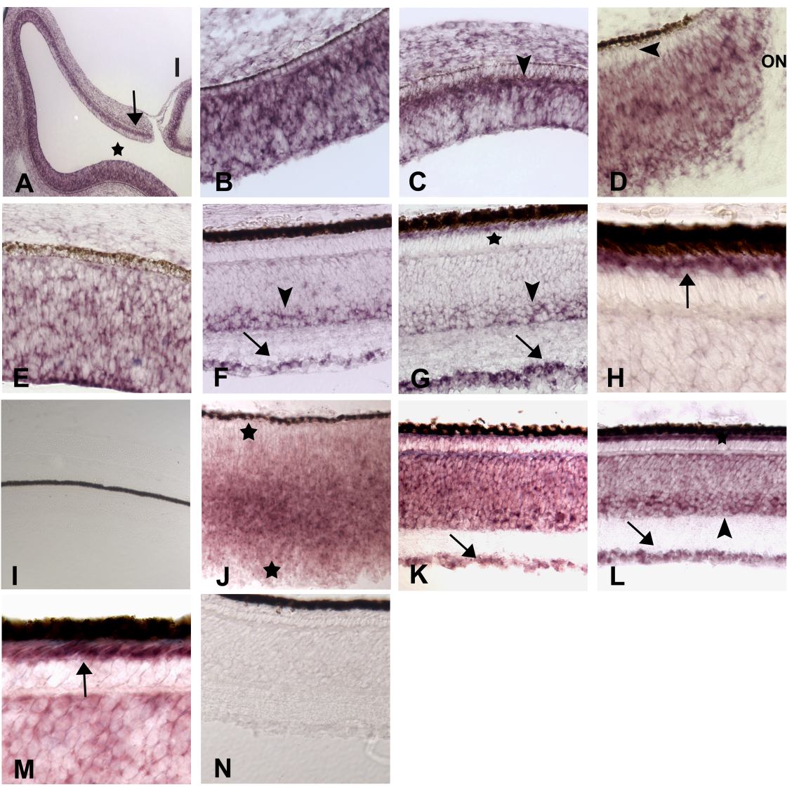
Figure 2. Developmentally regulated expression patterns of novel chick homologs
A-I: CRG9, homologous to dystrophin-like human predicted protein KIAA0728. A-C: ED 5. A: Low power view of the retina and lens (l). Signals are widespread at the fundus (A*, B), but more restricted at the periphery (A, arrow, C). D: ED 8, fundus; the putative photoreceptor layer (arrowhead) is negative in the region adjacent to the optic nerve (ON), while the remaining neural retina appears very positive. E: ED 8, periphery; heterogeneous expression is seen in the retinal epithelium. F-H: ED 18. F, periphery, G-H, fundus. Cells in the GCL (arrows) and the amacrine layer (arrowheads) are positive throughout the retina and much weaker or absent in the rest of the INL. Photoreceptor cells appear negative at the periphery (F), but are very strong in the fundal region (G, *) as illustrated in more detail in panel H (arrow). J-N: CRG31, homologous to human hypothetical protein FLJ20013. J: ED 8. Widespread expression, predominating in the outer most regions of the neural epithelium (*). K: ED 15. GCL (arrow) and the INL have positive signals. L and M: ED 18. L: Ganglion cells (arrow), amacrine layer (arrowhead) and other cells scattered throughout the INL and in the photoreceptor layer (*) show strong signals. Photoreceptor signals can be seen at higher magnification to localize to the inner segment region of these cells (M, arrow). I and N: Sense control probes for CRG9 and CRG31, respectively.