![]() Figure 4 of
Reich, Mol Vis 2003;
9:210-216.
Figure 4 of
Reich, Mol Vis 2003;
9:210-216.
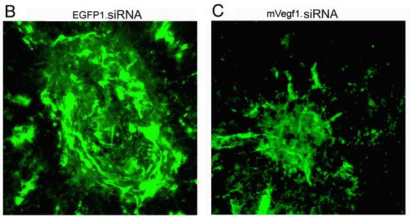
Figure 4. The extent of CNV is significantly reduced after subretinal delivery of mVegf1.siRNA
Thirty-six hours following laser photocoagulation, mVegf1.siRNA was delivered subretinally in one eye of each of 30 mice. Contralateral eyes were injected with a control (EGFP1) siRNA. Animals were perfused with dextran-fluorescein and the areas of CNV were measured in choroidal flat mounts 14 days after laser treatment. There is a significant difference (p<0.003) in mean areas of CNV between eyes injected with mVegf1.siRNA versus EGFP1.siRNA (panel A). Representative areas of CNV in eyes of a dextran-fluorescein-perfused animal that had received EGFP1.siRNA in one eye but mVegf1.siRNA in the other are shown in the colorized panels B and C, respectively. The CNV lesions were generally well-circumscribed by a region lacking fluorescence (as in panel B). CNV was identified by observing dextran-fluorescein-filled blood vessels on the choroidal/retinal interface, which are normally absent.