![]() Figure 3 of
Neethirajan, Mol Vis 2003;
9:205-209.
Figure 3 of
Neethirajan, Mol Vis 2003;
9:205-209.
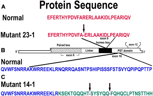
Figure 3. Comparison of normal and mutant protein sequences of the PAX6 gene
A: PAX6 exon 9 derived from proband 23-1 shows the premature truncation of protein with stop codon (arrow). B: Graphical representation of the PAX6 gene showing the paired box, homeo box, glycine rich region, and PST domain. C: PAX6 exon 10 derived proband 14-1 shows the frameshift mutation which leads to premature truncation of protein by a stop codon (arrow).
