![]() Figure 6 of
Dentchev, Mol Vis 2003;
9:184-190.
Figure 6 of
Dentchev, Mol Vis 2003;
9:184-190.
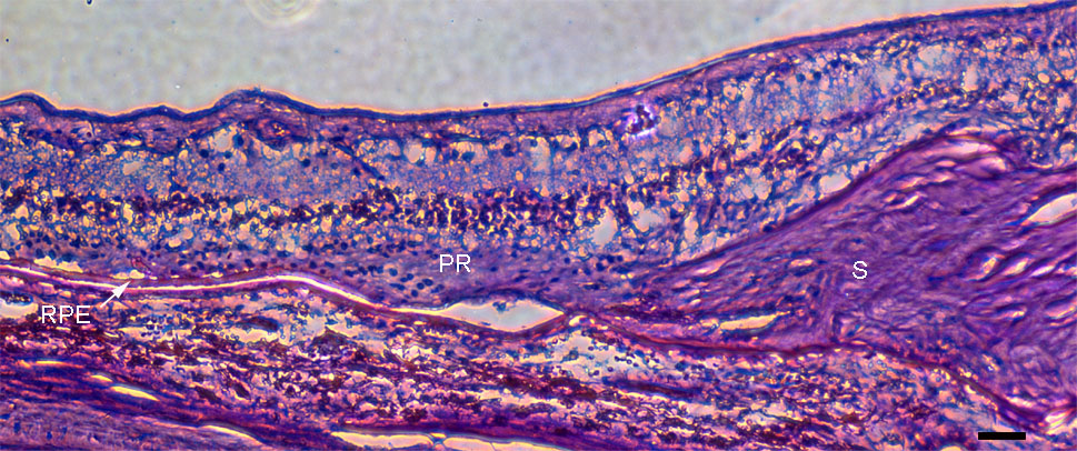
Figure 6. Exudative AMD retina 00-17
Photomicrograph of exudative AMD retina 00-17 stained with PAS-hematoxylin. This retina had no Aβ-positive vesicles on ICC (not shown). Thick exudative scar (S) is present on the right. Photoreceptors (PR) are present just temporal (to the left) to scar. Further temporal, the RPE monolayer is present, with minimal sub-RPE deposit. Scale bar represents 50 μm.