![]() Figure 5 of
Dentchev, Mol Vis 2003;
9:184-190.
Figure 5 of
Dentchev, Mol Vis 2003;
9:184-190.
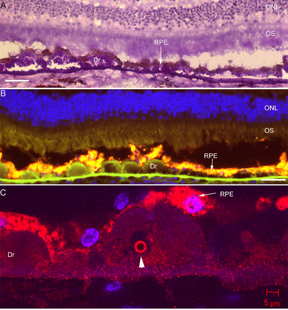
Figure 5. Few Aβ positive drusen in early AMD retina 00-1676
Photomicrographs showing Aβ in only a few areas in early AMD retina 00-1676. A: PAS-hematoxylin-stained low power image with soft drusen and minimal photoreceptor atrophy. B: Anti-Aβ antibody 6E10 is negative in this section. C: In another area, anti-Aβ antibody 2332 detects a single vesicle and punctate granular deposits within drusen. Image was acquired by confocal microscopy. Scale bar represents 50 μm.