![]() Figure 4 of
Dentchev, Mol Vis 2003;
9:184-190.
Figure 4 of
Dentchev, Mol Vis 2003;
9:184-190.
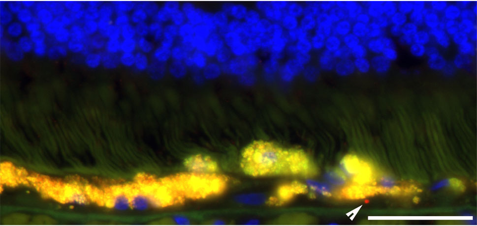
Figure 4. Single Aβ positive vesicle in early AMD retina 00-32
Fluorescent photomicrograph showing Aβ in drusen in early AMD retina 00-32. Arrowhead indicates single Aβ-positive vesicle underlying RPE atrophy and displacement into the subretinal space. Scale bar represents 50 μm.