![]() Figure 2 of
Dentchev, Mol Vis 2003;
9:184-190.
Figure 2 of
Dentchev, Mol Vis 2003;
9:184-190.
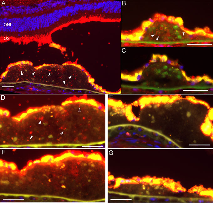
Figure 2. Aβ in drusen in GA retina 00-11
Fluorescent photomicrographs showing Aβ in drusen in GA retina 00-11. This donor had GA throughout much of the macula. A: Anti-Aβ mAb 4G8 label (red) is present in vesicles (arrowheads) within a large druse and photoreceptor outer segments at the temporal edge of atrophy. Nuclei are labeled with DAPI (blue). RPE cytoplasm appears gold due to lipofuscin autofluorescence. ONL: outer nuclear layer; OS: outer segments. B: Anti-Aβ pAb 2332 labels vesicles (arrowheads) within another large druse. C: Pre-adsorption with Aβ peptide eliminates label. D-G: are closely spaced sections labeled with anti-Aβ mAb4G8 (D), secondary antibody only (E), anti-rhodopsin antibody mAb 4D2 (F), and 4G8 pre-adsorbed with Aβ peptide (G). Of these four images, the only one with significant Aβ-positive vesicles is (D), verifying labeling specificity. Scale bar represents 50 μm.