![]() Figure 2 of
Burt, Mol Vis 2003;
9:164-170.
Figure 2 of
Burt, Mol Vis 2003;
9:164-170.
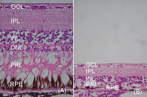
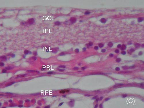
Figure 2. Comparison of the pathology of normal and rdd chicken retinas at 3-4 months
Comparison of normal (A) and rdd (B) retina at 3-4 months of age, both to scale. The rdd retina shows marked atrophy of all layers with virtually complete loss of photoreceptors and replacement by gliosis. GCL = ganglion cell layer, IPL = inner plexiform layer, INL = inner nuclear layer, PRL = photoreceptor layer and RPE = retinal pigment epithelium. Both haematoxylin and eosin at 340x magnification. (C) High power view of the rdd retina showing marked atrophy. Note in particular the complete absence of the outer nuclear layer (usually seen between the inner nuclear and photoreceptor layers). The photoreceptor layer shows loss of photoreceptors, gliosis and prominent clefts above the remnant of the retinal pigment epithelium. GCL = ganglion cell layer, IPL = inner plexiform layer, INL = inner nuclear layer, PRL = photoreceptor layer, RPE = retinal pigment epithelium. Haematoxylin and eosin at 1,800x magnification.