![]() Figure 4 of
Seigel, Mol Vis 2003;
9:159-163.
Figure 4 of
Seigel, Mol Vis 2003;
9:159-163.
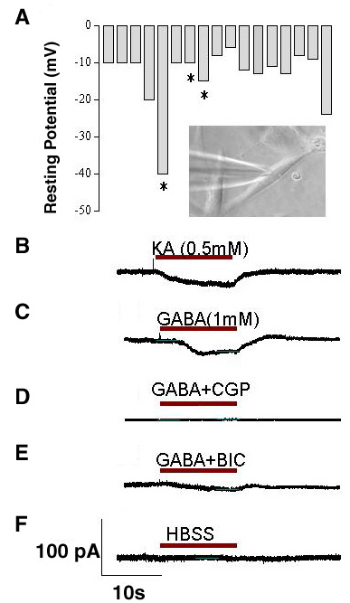
Figure 4. Electrophysiology of corneal stem cells in vitro
A: Resting potentials of corneal stem cells measured in current clamp were relatively low (mean±SD: -13±8 mV, n=13; range -6 mV to -40 mV). No action potentials or voltage sensitive Na+ or K+ currents were detected. Asterisks indicate cells in which KA or GABA evoked a response. Inset shows patch pipette attached to corneal stem cell. B: Kainic acid (KA, 0.5 mM), a non-NMDA glutamate receptor agonist, and C: GABA (1 mM) induced a small inward currents in a small number of corneal stem cells. D: The GABAb receptor antagonist, CGP-35348 (0.5 mM), and E: the GABAa receptor antagonist, bicuculline (BIC, 0.5 mM) blocked the GABA response. F: Puffing Hank's balanced salt solution (HBSS) onto corneal stem cells failed to induce a response. Horizontal bar shows duration of receptor agonists, antagonists, and control solution in B-F.