![]() Figure 3 of
Seigel, Mol Vis 2003;
9:159-163.
Figure 3 of
Seigel, Mol Vis 2003;
9:159-163.
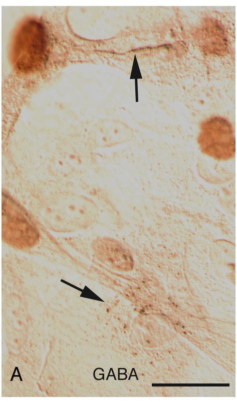
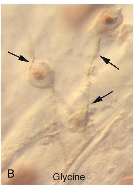
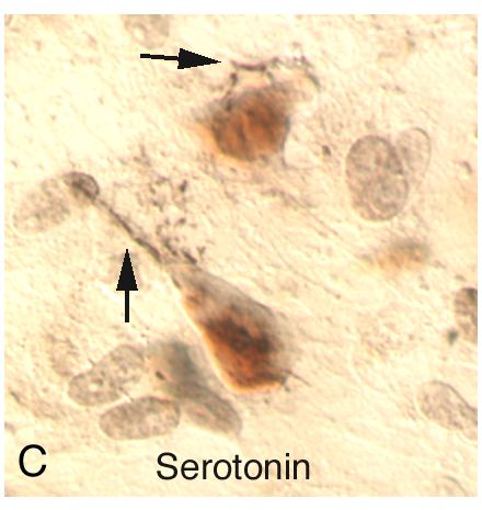
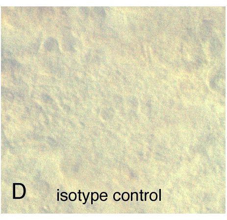
Figure 3. Corneal stem cells exhibit immunoreactivity for neurotransmitter receptors
Co-expression of the p63 corneal stem cell marker (red nuclei) with A: GABA receptor (note brown ridge (arrow) and "buckshot" pattern (arrowhead)); B: Glycine receptor (arrows); C: Serotonin receptor (arrows). D: Rabbit anti-dopamine receptor immunonegativity shown as isotype control. Scale bar represents 5 μm.