![]() Figure 4 of
Chen, Mol Vis 2003;
9:151-158.
Figure 4 of
Chen, Mol Vis 2003;
9:151-158.
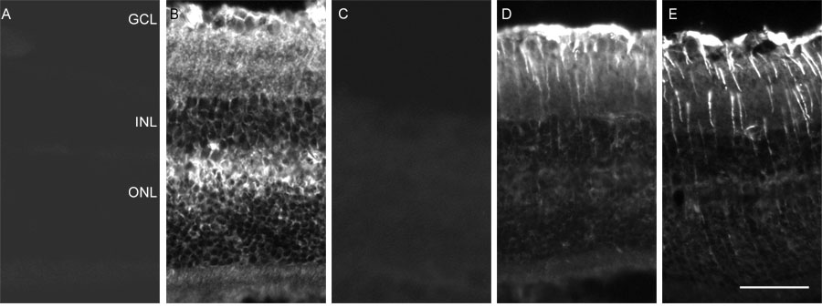
Figure 4. Ceruloplasmin IHC label in mouse retina
Immunohistochemistry with ceruloplasmin antibody demonstrates label throughout a normal retina, with increased label following photic injury. Fluorescence photomicrographs of mouse retinas labeled with no primary antibody (A), anti-ceruloplasmin (B,C), or anti-GFAP (D,E). A-C were normal retinas. D and E were 28 h after the end of photic injury (t28). Primary antibody in C and E was pre-adsorbed with ceruloplasmin protein before it was applied to the section.