![]() Figure 1 of
Chen, Mol Vis 2003;
9:151-158.
Figure 1 of
Chen, Mol Vis 2003;
9:151-158.
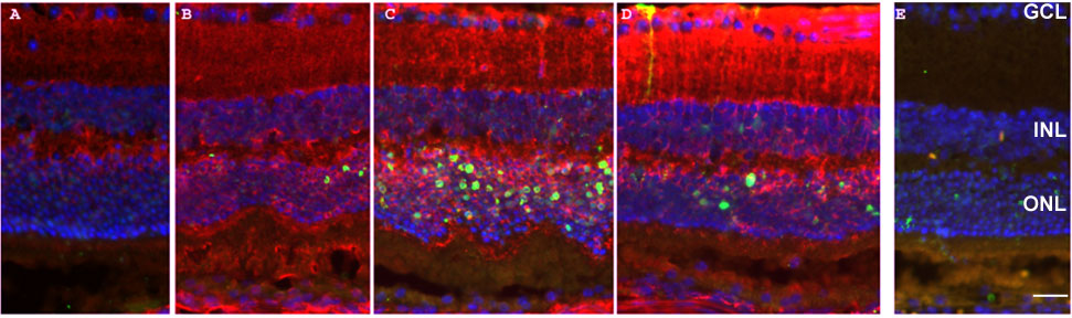
Figure 1. Photic injury induces TUNEL positive photoreceptors and elevated ceruloplasmin
Fluorescence photomicrographs of TUNEL (green) and anti-ceruloplasmin (red) labeled retinas at various times following photic injury. A: No light control. B: Immediately after photic injury (t0). C: Eight hours after photic injury (t8). D: Twenty eight hours after photic injury (t28). E: No primary ceruloplasmin antibody. Nuclei are labeled with DAPI (blue). Ganglion cell layer (GCL), inner nuclear layer (INL), outer nuclear layer (ONL). Scale bar 50 μm.