![]() Figure 4 of
Bode, Mol Vis 2003;
9:144-150.
Figure 4 of
Bode, Mol Vis 2003;
9:144-150.
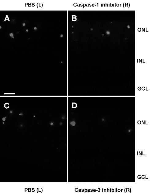
Figure 4. Apoptotic photoreceptor cells in retinas of tubby mice after injection of caspase inhibitors
TUNEL-stained longitudinal section through retinas of a PN 19 tubby mouse, previously injected with physiological saline (left eye control; A) and caspase-1 inhibitor (right eye; B). Between the control and the caspase-1 treated retina no difference in the number of apoptotic nuclei were visible. TUNEL-stained longitudinal section through the retinas of a PN 19 tubby mouse, previously injected with physiological saline (left eye control; C) and caspase-3 inhibitor (right eye). Comparison of both retinas revealed an obvious reduction of TUNEL-positive (D). INL, inner nuclear layer; GCL, ganglion cell layer; L, left eye; R, right eye. Scale bar represents 10 μm.