![]() Figure 3 of
Bode, Mol Vis 2003;
9:144-150.
Figure 3 of
Bode, Mol Vis 2003;
9:144-150.
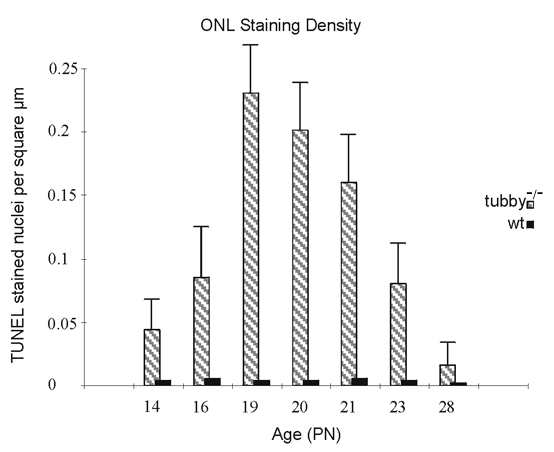
Figure 3. Time course of apoptotic photoreceptor cells of tubby and wild type mice
Retinas were collected at PN 14-28 from wild type and tubby mice. The number of TUNEL-positive apopototic photoreceptor cells in tubby mouse retinas increased after retinal differentation and passed an peak ("apoptotic peak"), before dropping to a small basic number. For each time point, the left, stripped column represents the number of TUNEL-positive nuclei per square μm of the outer nuclear layer in tubby mice. The right, black column represents that of wild type mice. The data of each time point represent the mean of counts of three representative areas of two retina samples; the error bars represent the standard error of the mean.