![]() Figure 1 of
Bode, Mol Vis 2003;
9:144-150.
Figure 1 of
Bode, Mol Vis 2003;
9:144-150.
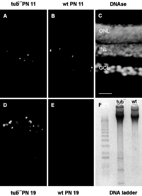
Figure 1. Apoptotic cells during the development of the retina in tubby and wild type mice
TUNEL-staining of longitudinal sections through retinas of PN 11 tubby mouse (A), PN 11 wild type mouse (B), control adult wild type retina treated with DNAse (C), PN 19 tubby mice (D) and PN 19 wild type mouse (E). In PN 11 TUNEL-positive apoptotic cells were present in the inner retina of both wild type and tubby mouse. In contrast, in PN 19 mice TUNEL-labeled apoptotic cells were restricted to the tubby retina where they localize to the outer nuclear layer (ONL). As expected, in the control DNAse-treated retina all nuclear layers were TUNEL-positive. INL, inner nuclear layer GCL, ganglion cell layer. Scale bar in C represents 20 μm. F: DNA ladder formation after agarose gel electrophoresis of genomic DNA confirmed apoptosis in PN 19 tubby mouse retinas (lane 1: DNA marker; lane 2: DNA ladder of PN 19 tubby mouse tub-/-; lane 3: no DNA ladder of PN 19 wild type mouse (wt)).