![]() Figure 1 of
van Lith-Verhoeven, Mol Vis 2003;
9:138-143.
Figure 1 of
van Lith-Verhoeven, Mol Vis 2003;
9:138-143.
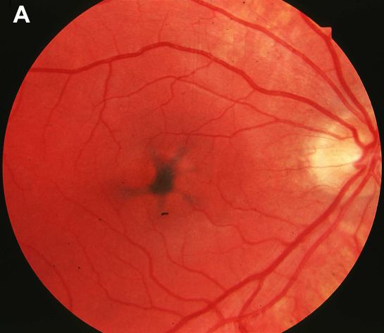
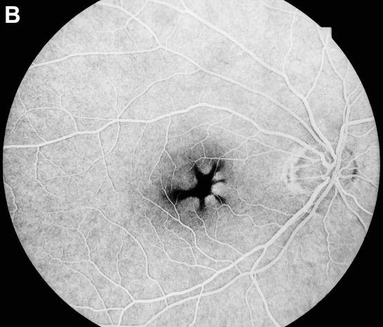
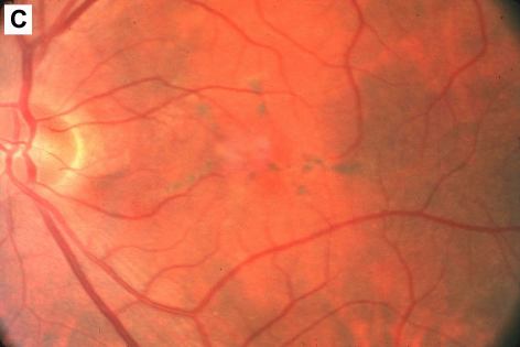
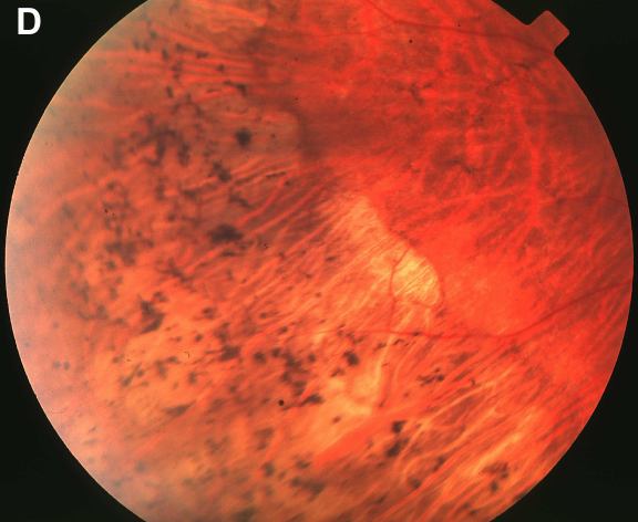
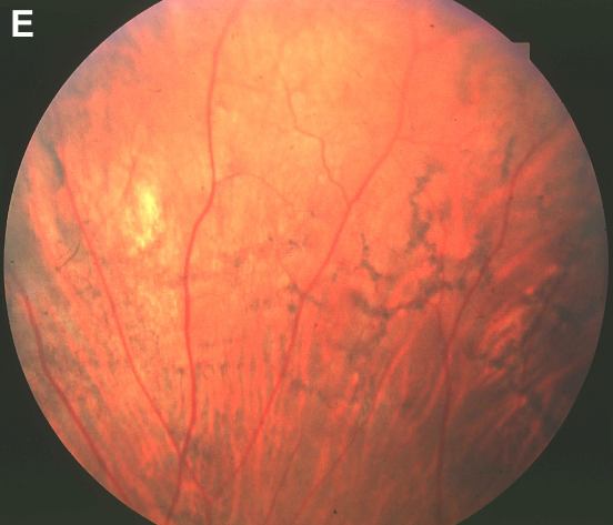
Figure 1. Fundus photographs and fluorescence angiography of affected BSMD family members
A: Fundus photograph of patient III-5 at age 16 years showing a black butterfly-shaped structure. B: Fluorescence angiography of individual III-5, showing a striking pigmented structure in front of a brightly fluorescent choroid. C: Fundus photograph of patient II-4 at age 52 years showing a more or less butterfly-shaped, slightly pigmented structure. D: Peripheral bone spicule-like structures and some parafoveal chorioretinal atrophy in patient II-4. E: Peripheral reticular pigmentations in patient II-3.