![]() Figure 1 of
Mawrin, Mol Vis 2003;
9:10-13.
Figure 1 of
Mawrin, Mol Vis 2003;
9:10-13.
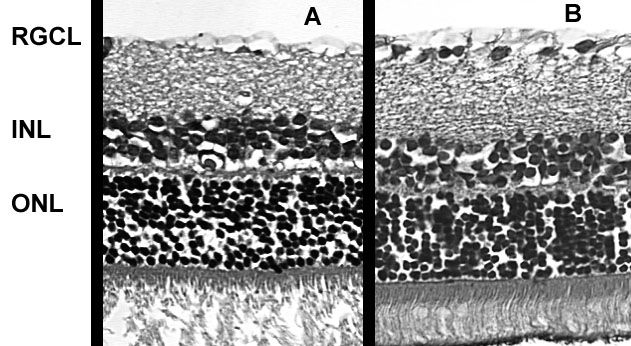
Figure 1. RGC loss in retinal cross sections after ONC
Hematoxilin-eosin stain of a representative cross sections of a retina 14 days post-ONC (A) and a sham-operated control retina (B). A marked reduction in retinal ganglion cells is evident. The retinal ganglion cell layer (RGCL), inner nuclear layer (INL), and outer nuclear layer (ONL) are labeled.