![]() Figure 4 of
Bowne, Mol Vis 2003;
9:129-137.
Figure 4 of
Bowne, Mol Vis 2003;
9:129-137.
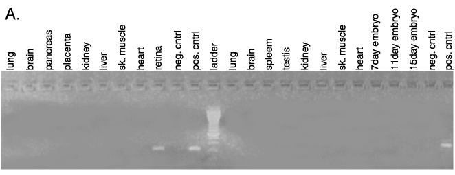
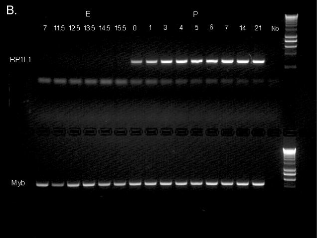
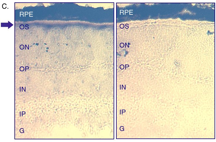
Figure 4. Expression analyses of RP1L1
A: PCR analyses of RP1L1 expression in mouse and human tissues. Human tissues are to the left of the molecular weight ladder in lanes 1-11 and mouse tissues are to the right in lanes 13-25. B: Pattern of RP1L1 expression in different aged mouse retinas. RP1L1 is found in the retinas of mice at birth, but does not appear before embryonic day 15.5. The control probe is mybP42POP. The band underneath RP1L1 is primer-dimer and the last lane is marker. P: postnatal day; E: embryonic day; NO is no DNA negative control. C: In situ hybridization of mouse retina with RP1L1 antisense (right) and sense (left) probes. Arrow shows hybridization of antisense probe in inner segments of photoreceptors.