![]() Figure 5 of
Al-Ghoul, Mol Vis 2003;
9:119-128.
Figure 5 of
Al-Ghoul, Mol Vis 2003;
9:119-128.
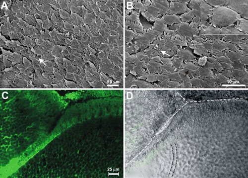
Figure 5. The peri-sutural region
A and B: SEM micrographs of intact fiber ends. Paired fluorescent (C) and differential interference contrast (D) images from a whole-mount specimen. In this region, fiber ends were oriented in curved or straight rows (one curved row is delineated by black dotted lines) at angles to the posterior pole and approaching posterior suture branches (white dotted lines). Ultrastrucural examination of intact fiber ends revealed that many ends possessed microspikes that extended in the direction of migration (black arrows in B and inset of B). White arrows indicate the direction of fiber end migration.