![]() Figure 3 of
Al-Ghoul, Mol Vis 2003;
9:119-128.
Figure 3 of
Al-Ghoul, Mol Vis 2003;
9:119-128.
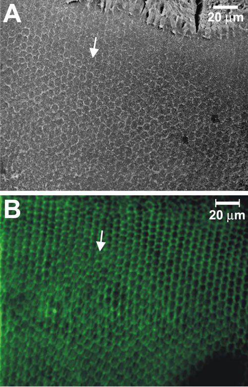
Figure 3. The equatorial region
A: SEM micrograph of a whole-mount lens capsule. In this region basal ends were hexagonal in shape and arranged in rows oriented toward the posterior pole. B: LSCM micrograph of a phalloidin-treated whole-mount lens capsule. F-actin fluorescence in the BMC showed a comparable pattern of shape and arrangement to that seen in SEMs, indicating that F-actin delineated the extent of the BMC in posterior ends. Arrows indicate the direction of migration.