![]() Figure 2 of
Al-Ghoul, Mol Vis 2003;
9:119-128.
Figure 2 of
Al-Ghoul, Mol Vis 2003;
9:119-128.
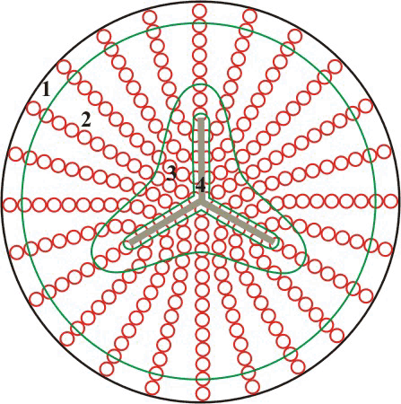
Figure 2. Diagram of migrating posterior fiber ends adhered to the capsule
Morphometry and morphology of the basal fiber ends was assessed within 4 regions, demarcated by green lines and designated the equatorial region, the lateral-posterior region, the peri-sutural region, and the sutural region. Fiber ends are red and sutures are gray.