![]() Figure 1 of
Al-Ghoul, Mol Vis 2003;
9:119-128.
Figure 1 of
Al-Ghoul, Mol Vis 2003;
9:119-128.
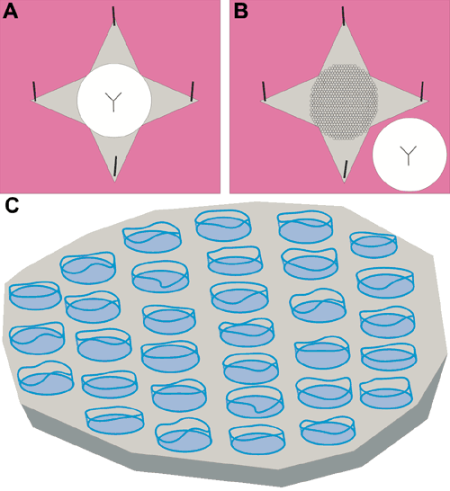
Figure 1. Diagrammatic representation of the technique for preparation of whole-mount lens capsules
A: After light prefixation, lenses were washed in buffer, placed posterior side down on squares of dental wax, and the capsule was carefully peeled away from the fiber mass and pinned down to the wax (gray=capsule; white=fiber mass; pink=dental wax). B: The lens mass was lifted away from the capsule and discarded, then the whole-mount capsule with adhered fiber ends (honeycomb pattern) was fixed and processed for either SEM or LSCM. C: Enlarged view of a portion of a whole mount lens capsule. This technique provided specimens in which the posterior ends of fibers were broken away from the fiber mass (irregular flattened cylinders) and adhered to the capsule (gray) via the BMC (blue) of each fiber. Fiber ends are shown spaced apart in the diagram for clarity only; they were directly adjacent to one another on actual specimens.
