![]() Figure 5 of
Srivastava, Mol Vis 2003;
9:110-118.
Figure 5 of
Srivastava, Mol Vis 2003;
9:110-118.
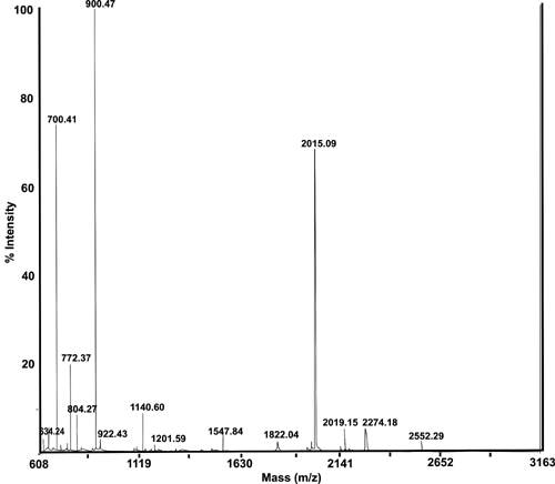
Figure 5. MALDI-TOF profile of tryptic fragments of spot number 1
MALDI-TOF spectrum of spot number 1 following trypsin digestion. The spectrum represented a typical mass spectrum of tryptic fragments from spots 1-5 from WS proteins and spots number 9-13 from WI proteins of normal lenses (see Figure 2A,B). The MALDI-TOF spectra of tryptic fragments from spots number 1-14, isolated from WS and WI proteins of cataractous lenses (see Figure 3) were similar to the typical spectrum shown.