![]() Figure 2 of
Fernandez-Durango, Mol Vis 2003;
9:103-109.
Figure 2 of
Fernandez-Durango, Mol Vis 2003;
9:103-109.
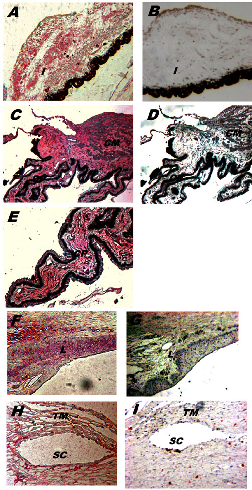
Figure 2. Localization of ET-1 immunoreactivity in the anterior segment of the human eye
Immunolocalization of ET-1 (A, C, E, F, H). Positive ET-1 immunoreactivities (red) were shown in the iris (A), the CM (C), the stroma and NPCE cells of the CP (C, E), the epithelial cells of limbus (F), the trabecular cells and in the outer and inner walls of Schlemm's canal (H). Negative controls were obtained with anti-ET-1 preabsorbed with 10 nM ET-1 (B, D, G, I). All images were magnified 66 times. Abbreviations are used in the figure for iris (I), ciliary process (CP), ciliary muscle (CM), limbus (L), trabecular meshwork (TM), and Schlemm's canal (SC).