![]() Figure 3 of
Bourcier, Mol Vis 2003;
9:96-102.
Figure 3 of
Bourcier, Mol Vis 2003;
9:96-102.
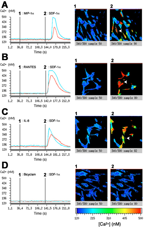
Figure 3. Effect of various chemokines on intracellular calcium mobilization in cultured corneal fibroblasts
When corneal fibroblasts were triggered with 100 nM MIP-1 (A1), RANTES (B1) or IL-8 (C1), no increase in the intracellular calcium concentration was observed. Conversely, when 10 nM SDF-1 was delivered (A2,B2,C2), a significant response within few seconds was detected. The addition of 1 μM bicyclam (D1) before stimulation with SDF-1 (D2) significantly inhibited the calcium response. Images ratio of Fura-2 after application of chemokines or bicyclam (1) and SDF-1 (2) represent one typical experiment. The red line corresponds to the mean variation of intracellular calcium for the whole field. The blue line presented the calcium evolution in a typical reactive corneal fibroblast outlined by a small circle within the photos (arrowheads).