![]() Figure 2 of
Hagan, Mol Vis 2003;
9:87-92.
Figure 2 of
Hagan, Mol Vis 2003;
9:87-92.
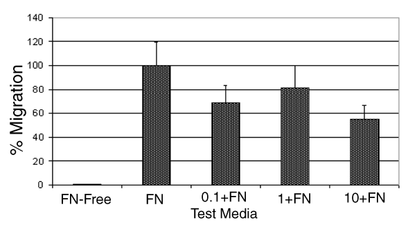
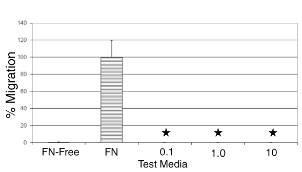
Figure 2. Effects of SPARC on HRPE and FN-induced HRPE cell migration
The migratory behavior of HRPE cells was assessed in standard 48-well microchemoattraction (Boyden) chamber experiments. The results are expressed as a percentage of the positive control (10 μg/ml fibronectin in F10 media), which is set at 100%. A: Test media: "FN-free" was F10 media only (negative control); "FN" was fibronectin at 10 μg/ml in F10 media (positive control); "0.1", "1.0", and "10" were the respective concentrations of SPARC (μg/ml) as indicated in the figure, diluted in F10 media only. No migration towards SPARC was observed for any of the concentrations used and this was found to be significantly different from the FN positive controls (*p<0.001). B: Test media: "FN-free" was F10 media only (negative control); "FN" was fibronectin at 10 μg/ml in F10 media (positive control); "0.1+FN", "1+FN", and "10+FN" were the respective concentrations of SPARC (μg/ml) as indicated in the figure, in the presence of 10 μg/ml FN in F10 media. Migration towards a source of FN was diminished with all concentrations of SPARC used, however, these results did not reach significance. Error bars represent the standard error of the mean.
A:
B: