![]() Figure 4 of
Armstrong, Mol Vis 2003;
9:74-79.
Figure 4 of
Armstrong, Mol Vis 2003;
9:74-79.
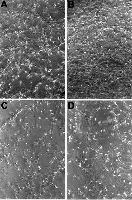
Figure 4. The effect of thrombospondin antibodies on the morphology of keratocytes cultured in collagen
Phase contrast micrographs of human corneal keratocytes in 3D collagen matrices showing cell morphology. A: Day 3, keratoctyes with untreated culture media. The cells are extending small processes into the surrounding collagen. B: Day 7, keratoctyes with untreated culture media. The cells forming contacts and reforming the collagen matrix. C: Day 3, keratoctyes with culture medium containing 20 μg/ml of TSP-2 antibody. The cells are predominantly rounded with only short, fine processes. D: Day 7, keratoctye populated collagen matrix in culture medium containing 20 μg/ml of TSP-2 antibody. The cells are still rounded, with some more processes, but are very different to the network seen in B.