![]() Figure 3 of
Armstrong, Mol Vis 2003;
9:74-79.
Figure 3 of
Armstrong, Mol Vis 2003;
9:74-79.
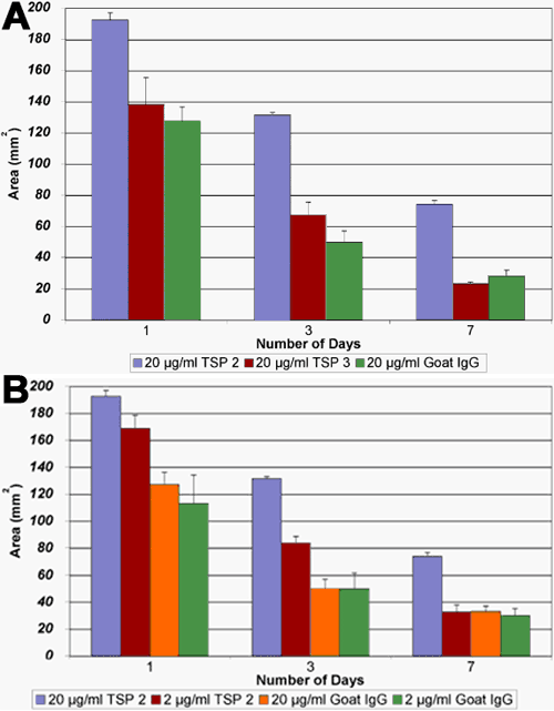
Figure 3. The effect of thrombospondin antibodies on keratocyte-mediated matrix contraction
A: Histograms showing the mean matrix area (error bars represent the standard deviation) for human corneal keratocyte-seeded (3D) collagen matrices in the presence or absence of antibodies directed against TSP-2 and TSP-3 (both antibodies at final concentration of 20 μg/ml). At days 1, 3, and 7 there were significant differences (p<0.01; n=9-11) between the TSP-2 antibody treated group and the untreated control group (20 μg/ml goat IgG). There was no significant difference between the control group and TSP 3 antibody treated matrices. B: Histograms showing the mean matrix area (error bars represent the standard deviation) for human corneal keratocyte-seeded (3D) collagen matrices in the presence or absence of antibodies directed against TSP-2 (antibodies at final concentration of 20 μg/ml and 2 μg/ml). At days 1, 3, and 7 there were significant differences (p<0.01; n=9-11) between the TSP-2 20 μg/ml antibody treated group and the untreated control group (20 μg/ml goat IgG). At days 1 and 3 there were significant differences (p<0.01; n=9-11) between the TSP-2, 2 μg/ml antibody treated group and the untreated control group (2 μg/ml goat IgG). On day 7 there was no significant difference between the control group (2 μg/ml goat IgG) and the TSP-2, 2 μg/ml antibody treated group.