![]() Figure 8 of
Perkins, Mol Vis 2003;
9:60-73.
Figure 8 of
Perkins, Mol Vis 2003;
9:60-73.
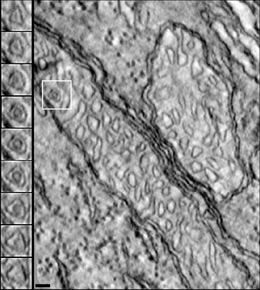
Figure 8. Unusual topography of a small matrix area completely surrounded by a crista
Because the matrix space is entirely bounded by a crista membrane, it is separated topographically from the mitochondrial periphery. This anomaly may be a structural defect or a viable, functioning unit. The multiple insets on the left show an enlarged area from serial slices through the 3-D reconstruction of the boxed region of the cone mitochondrion shown on the right. Scale bar represents 50 nm.