![]() Figure 3 of
Perkins, Mol Vis 2003;
9:60-73.
Figure 3 of
Perkins, Mol Vis 2003;
9:60-73.
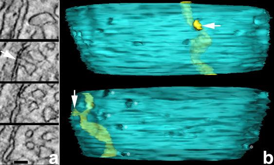
Figure 3. Perpendicular extensions connect tubular cristae
This figure demonstrates that tubular cristae in cone mitochondria connect to the inner boundary membrane through perpendicular extensions from the tubular shaft. A: Serial slices through the tomographic reconstruction of a cone mitochondrion. The serial slices illustrate how the crista membrane extends from the tubular shaft towards the periphery of the mitochondrion, forms a crista junction (arrow) and finally contracts away from the boundary membrane. Scale bar represents 50 nm. B: Volume segmentation and surface rendering of the same crista. The two 90° rotation views (top and bottom) highlight the dimensions of the crista junction opening (arrows) in relation to the crista (yellow) and mitochondrial inner membrane (blue). Crista junctions (*) from other cristae also are shown and are indicated by fenestrations in the inner membrane. These fenestrations accurately portray the size and variation in dimensions of these junctions.