![]() Figure 1 of
Perkins, Mol Vis 2003;
9:60-73.
Figure 1 of
Perkins, Mol Vis 2003;
9:60-73.
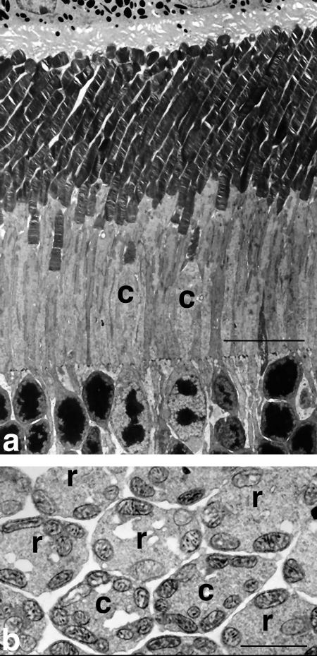
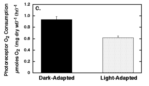
Figure 1. Electron microscopy and photoreceptor oxygen consumption of 21 day old mouse retina
Conventional electron micrographs of retinas and photoreceptor oxygen consumption (QOPR) from postnatal day 21 mice. A: Electron micrographs of the rod (r) and cone (c) outer and inner segments and distal outer nuclear layer from superior temporal retina of a light-adapted mouse. Note that the tips of the middle wavelength-sensitive cone outer segments lie in the rod inner segment region. The cone inner segment diameter is approximately twice that of the rod inner segment. The cone nuclei are located in the distal portion of the outer nuclear layer and contain several clumps of irregularly shaped heterochromatin, whereas the rod nuclei contain a single compact mass of heterochromatin. Scale bar represents 10 μm. B: Transverse section through ellipsoid region of photoreceptor inner segment illustrating cytochrome oxidase stained rod (r) and cone (c) mitochondria. Note that there are approximately twice as many cone mitochondria per photoreceptor compared to rod photoreceptors and that the cone mitochondrial inner membranes stain more intensely for cytochrome oxidase than do the rods. Scale bar represents 1 μm. C: QOPR. QOPR was measured in pairs of dark-adapted and rod saturating light-adapted whole retinas from five mice.