![]() Figure 1 of
Schulz, Mol Vis 2002;
8:67-71.
Figure 1 of
Schulz, Mol Vis 2002;
8:67-71.
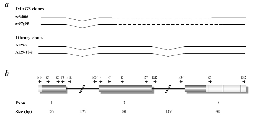
Figure 1. cDNA cloning and genomic structure of C7orf9
A: A consensus cDNA sequence was assembled from I.M.A.G.E. retina clones ze34f06 and ze37g05 and cDNA clones retrieved from cDNA library screenings. The dotted line indicates unknown sequence. B: C7orf9 comprises three coding exons which are represented by shaded boxes. The exon and intron sizes are derived from alignment of the cDNA to the corresponding human draft sequence. Light gray boxes represent the 5'-and 3'-untranslated regions (UTRs). Three potential poly(A)adenylation sites are depicted as vertical black bars in the 3'-UTR. The name and relative positions of oligonucleotide primers utilized in the study are shown.