![]() Figure 6 of
Liou, Mol Vis 2002;
8:494-501.
Figure 6 of
Liou, Mol Vis 2002;
8:494-501.
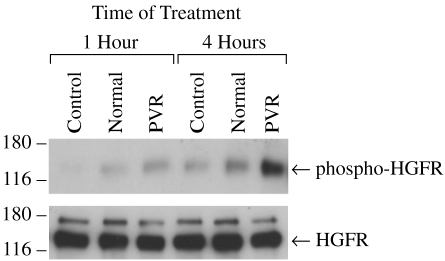
Figure 6. Tyrosine phosphorylation of HGFR in ARPE-19 cells treated with rabbit vitreous
Serum starved subconfluent ARPE-19 cells were treated for 1 or 4 h with vitreous humor from eyes 24 h after surgery or with vitreous from normal eyes. Protein equivalent aliquots of cell lysates were immunoprecipitated with anti-phosphotyrosine antibody and then analyzed by western blot using anti-HGFR antibody (top) or directly analyzed by western blot using anti-HGFR antibody (bottom). The positions and the molecular weight (kD) of protein markers are indicated.