![]() Figure 4 of
Liou, Mol Vis 2002;
8:494-501.
Figure 4 of
Liou, Mol Vis 2002;
8:494-501.
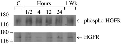
Figure 4. Tyrosine phosphorylation of c-met induced by the retinal holes and inflammation
Immuno-precipitation and immuno-blot analysis of HGFR (c-met) activation in retinal and RPE cells from normal eyes and eyes with retinal holes produced by surgery followed by IL-1β injections. Protein equivalent aliquots of retinal and RPE lysates collected from normal eyes or from post-surgical eyes at different post-surgical time points were immunoprecipitated with anti-phosphotyrosine antibody and then analyzed by western blot using anti-HGFR antibody (top) or directly analyzed by western blot using anti-HGFR antibody (bottom). The positions and the molecular weight (kD) of protein markers are indicated.