![]() Figure 2 of
Liou, Mol Vis 2002;
8:494-501.
Figure 2 of
Liou, Mol Vis 2002;
8:494-501.
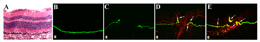
Figure 2. Proliferative RPE cells around the retinal holes with inflammation
Hematoxylin and eosin stained and anti-pan-cytokeratin (Oregon Green)/anti-PCNA (Texas Red) double stained sections of posterior segments in normal eyes and eyes with retinal holes produced by surgery followed by IL-1β injections. A: Normal eye, Hematoxin & Eosin stained; B: Normal eye, anti-pan-cytokeratin/anti-PCNA double-stained C: Post-surgically treated eye (72 h), anti-pan-cytokeratin/anti-PCNA double-stained. D,E: Post-surgically treated eye (4 weeks), anti-pan-cytokeratin/anti-PCNA double stained. Arrows indicate proliferating RPE cells.