![]() Figure 1 of
Liou, Mol Vis 2002;
8:494-501.
Figure 1 of
Liou, Mol Vis 2002;
8:494-501.
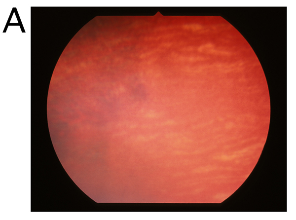
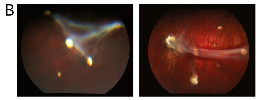
Figure 1. Fundus photographs of normal rabbit eyes and eyes with retinal holes and inflammation
Fundus photographs of normal rabbit eyes and eyes containing retinal holes produced by surgery followed by IL-1β injections. A: Normal eye. B: Surgically treated eyes (4 weeks post-surgery); membranes have formed over the holes and extended into the vitreous body, forming adhesions with other portions of the retina.