![]() Figure 9 of
Liou, Mol Vis 2002;
8:483-493.
Figure 9 of
Liou, Mol Vis 2002;
8:483-493.
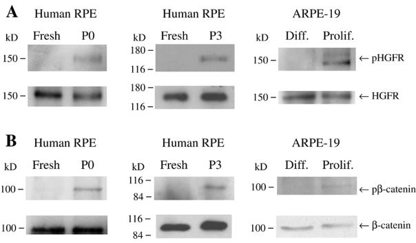
Figure 9. Tyrosine phosphorylation of HGFR and β-catenin
Tyrosine phosphorylation of HGFR (A) and β-catenin (B) in freshly isolated or subconfluent cultures of passaged or non-passaged RPE cells, or in differentiated or subconfluent ARPE-19 cells. Protein equivalent aliquots of tissue or cell lysates were immunoprecipitated with anti-HGFR or anti-β-catenin antibody and then analyzed by western blot with anti-phosphotyrosine antibody. The membrane was stripped and analyzed with anti-HGFR or anti-β-catenin antibody. pHGFR indicates phosphorylated HGFR and pβ-catenin indicates phosphorylated β-catenin. The positions and the molecular weight (kD) of protein markers are indicated.