![]() Figure 2 of
Liou, Mol Vis 2002;
8:483-493.
Figure 2 of
Liou, Mol Vis 2002;
8:483-493.
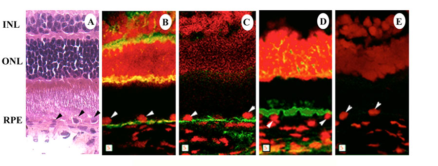
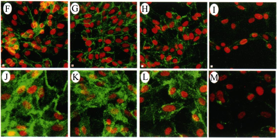
Figure 2. Laser scanning confocal microscopic immunolocalization of β-catenin
Laser scanning confocal microscopic immunolocalization of β-catenin in rabbit retina, in ARPE-19 cells, and in cultured human RPE cells. A-E: Rabbit retina. A: Hematoxylin and eosin stained cryosection depicting inner and outer nuclear layer (INL and ONL) and RPE cells. B-E: Vertical sections (x and z) taken of cryosection of rabbit retina incubated with different IgGs and with Oregon Green conjugated goat-anti-mouse IgG. B: anti-β-catenin; C: anti-E-cadherin; D: anti-Na+/K+ ATPase; E: normal mouse serum. Cell nuclei were stained (red) with propidium iodide. Arrowheads indicate nuclei of RPE cells. F-M: Effect of wounding and HGF induced migration of ARPE-19 cell (F-I) and cultured human RPE cells (J-M) on β-catenin re-distribution. Confluent cells on chamber slides were starved, scratched, and HGF added. After 8 h, the cells were fixed and labeled with anti-β-catenin IgG and Oregon Green conjugated goat-anti-mouse IgG. F,J: Untreated cells; G,K: HGF treated cells away from the wound; H,L: HGF treated cells closer to the wound; I,M: HGF treated cells at the wound. Squares represent 5 μm.