![]() Figure 6 of
Margulis, Mol Vis 2002;
8:477-482.
Figure 6 of
Margulis, Mol Vis 2002;
8:477-482.
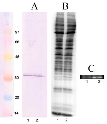
Figure 6. Cyclic GMP-dependent phosphorylation of phosducin in permeabilized nuclear preparation
Purified nuclear fraction was permeabilized by freezing and thawing, phosphorylated in the presence of γ-32PATP, electrophoresed, transferred to PVDF membrane, and probed with the anti-phosducin antibody. Panel A: Western blot; Panel B: Corresponding autoradiogram. Panel C: shows the negative of a part of an autoradiogram corresponding to the position of phosducin. This autoradiogram was from a longer exposure to the same gel and the insert shows the difference between the two lanes more clearly. Lane 1, control no additions; lane 2, 0.1 mM 8-Br-cyclic GMP. On the left of Panel A are shown standard proteins with their molecular weights marked in kDa.