![]() Figure 5 of
Margulis, Mol Vis 2002;
8:477-482.
Figure 5 of
Margulis, Mol Vis 2002;
8:477-482.
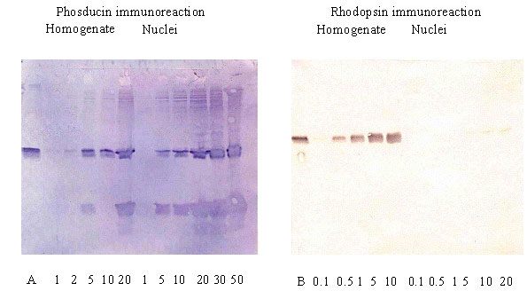
Figure 5. Estimation of rhodopsin and phosducin in retinal homogenate and purified nuclear preparation
Proteins of the retinal homogenate and purified nuclear preparation were subjected to electrophoresis, transferred to PVDF membrane and probed with anti-phosducin (left panel) or anti-rhodopsin (right panel) antibody. A: 100 ng of phosducin standard; B: 100 ng of rhodopsin standard. Numbers under lanes represent the amount of total protein loaded, in μg.