![]() Figure 3 of
Margulis, Mol Vis 2002;
8:477-482.
Figure 3 of
Margulis, Mol Vis 2002;
8:477-482.
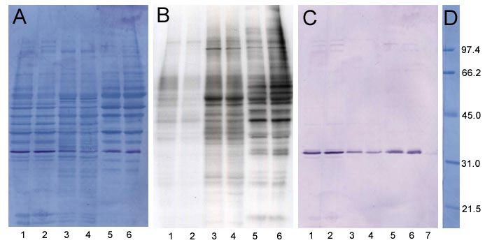
Figure 3. Phosphorylation of phosducin in subcellular fractions
After Phosphorylation in the presence of (γ-32P)ATP, homogenate was fractionated into 120x g pellet and 120x g supernatant. The supernatant was then centrifuged to obtain 100,000x g pellet and supernatant. The 120x g pellet was further processed to obtain purified nuclear fraction. Forty μg of protein from each fraction was electrophoresed, transferred to PVDF membrane, probed with an anti-phosducin antibody and autoradiographed. The western blot was then stained for protein with amidoblack. Panel A: Amidoblack staining for proteins of the western blot shown in Panel C; Panel B: Autoradiogram; Panel C: Western blot; Panel D: Amidoblack stained molecular weight markers. Lanes 1 and 2, purified nuclei; lanes 3 and 4, 100,000x g pellet membrane fraction; lanes 5 and 6, cytosolic fraction. Lanes 1, 3, and 5 are without additions; Lanes 2, 4, and 6 are from assays that contained 0.1 mM 8-Br-cyclic GMP. Lane 7, 100 ng of phosducin standard. Numbers in panel D mark molecular weight in kDa.