![]() Figure 2 of
Margulis, Mol Vis 2002;
8:477-482.
Figure 2 of
Margulis, Mol Vis 2002;
8:477-482.
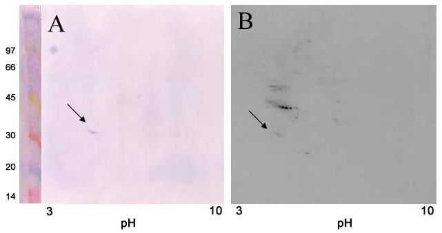
Figure 2. Identification of phosphorylated phosducin in cytosolic proteins after 2-dimensional separation
The cytosolic fraction phosphorylated in the presence of (γ-32P)ATP and 0.1 mM 8-Br-cyclic GMP was subjected to 2-D electrophoresis, and the separated proteins were transferred to PVDF membrane and probed with an anti-phosducin antibody. Panel A: Western blot; Panel B: Corresponding autoradiogram. Arrows identify phosducin. On the left of Panel A are shown standard proteins with their molecular weights marked in kDa. The abscissa shows the approximate pH of the first dimension separation. The reported pI of phosducin is 4.5 [19].