![]() Figure 1 of
Margulis, Mol Vis 2002;
8:477-482.
Figure 1 of
Margulis, Mol Vis 2002;
8:477-482.
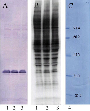
Figure 1. Cyclic nucleotide-dependent phosphorylation of phosducin in retinal homogenate
Proteins of bovine retinal homogenate were phosphorylated in the presence of (γ-32P)ATP, electrophoretically separated on a 10-20% polyacrylamide gradient gel, transferred to PVDF membrane and probed with an anti-phosducin antibody. A: Western blot, B: Corresponding autoradiogram, and C: Amidoblack stained molecular weight markers. Lane 1, control no additions; 2, 0.1 mM 8-Br-cyclic GMP; 3, 0.1 mM 8-Br-cyclic AMP; 4, standard proteins with molecular weights marked in kDa.